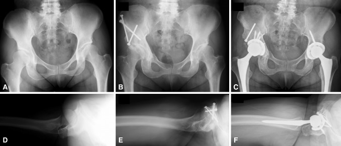
Fig. 1A–F.
(A) An anterior-to-posterior radiograph of the pelvis shows DDH with a 20° Tönnis angle and 10° lateral center-edge angle. (B) An anterior-to-posterior view of the pelvis after periacetabular osteotomy shows the acetabular correction with a 10° Tönnis angle and 25° lateral center-edge angle. (C) An anterior-to-posterior radiograph of the pelvis shows conversion to THA after periacetabular osteotomy on the patient’s right and without periacetabular osteotomy on the patient’s left. (D) A shoot-through lateral view of the hip shows acetabular anteversion. (E) A shoot-through lateral radiograph of the hip after periacetabular osteotomy shows overcorrection with acetabular retroversion. (F) A shoot-through lateral view of the hip shows conversion to THA with resection of the anterior wall and correction of the acetabulum to a more anteverted position.

