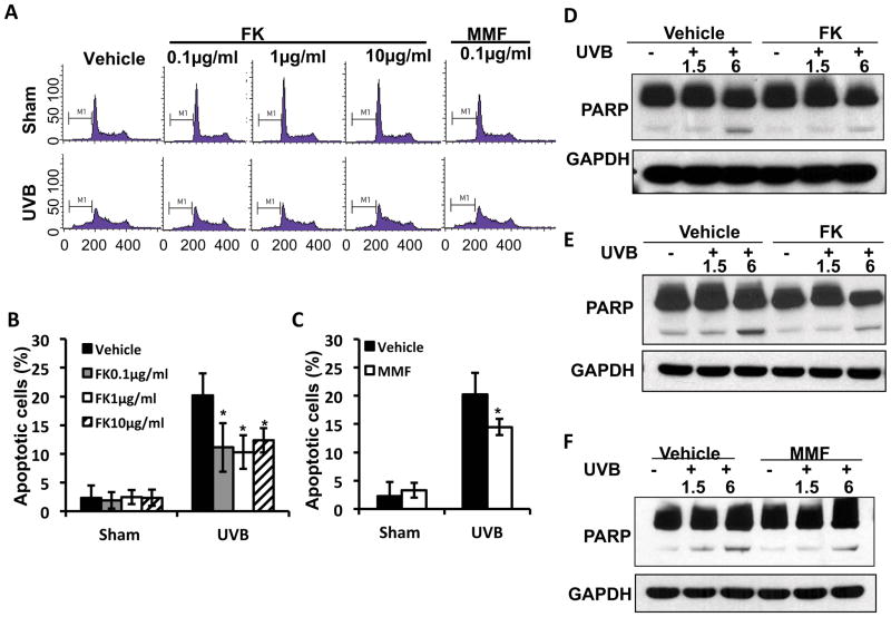
Figure 2. FK506 and MMF suppresses UVB-induced apoptosis in HaCaT cells.
(A) Flow cytometric analysis of UVB (30mJ/cm2)-induced apoptosis in HaCaT cells treated with vehicle, FK506 (0.1, 1 or 10 μg/ml) or MMF (0.1 μg/ml) for 1 week. (B–C) Quantitation of apoptotic cells (%) in A for FK506 treatment (B) and MMF treatment (C). *, P<0.05, t-test, significant differences between FK506 (0.1, 1 or 10 μg/ml), MMF (0.1 μg/ml) and vehicle control. (D–F) Immunoblot analysis of PARP and GAPDH in HaCaT cells at 1.5 or 6 h post-UVB (20mJ/cm2) after treatment with vehicle, FK (1 μg/ml) (D), FK (10 μg/ml) (E), or MMF (0.1 μg/ml) (F) for 1 week.

