
Figure 3. Heterosynaptic spine shrinkage requires close physical proximity to multiple potentiated spines.
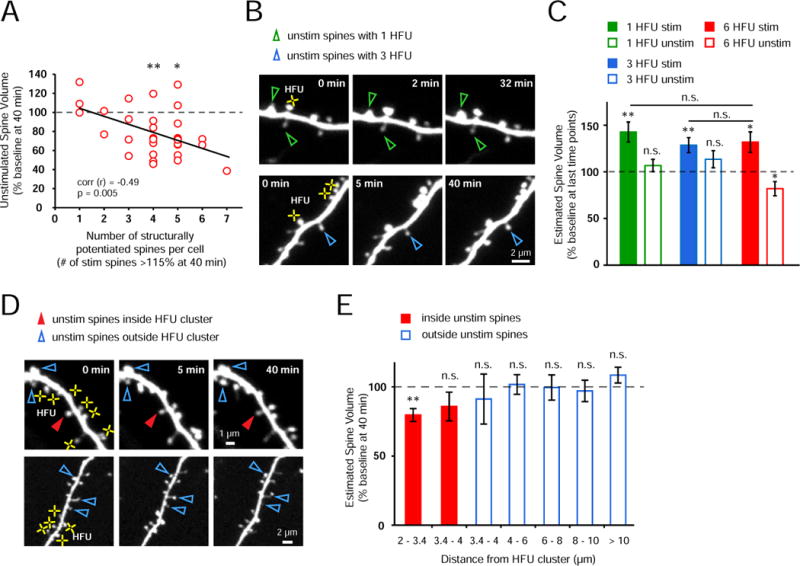
(A) Shrinkage of unstimulated spines was not observed on those cells for which HFU led to structural potentiation (> 115% of baseline at 40 min) of less than four spines; in contrast, when four or more spines potentiated, unstimulated spines shrank (4 spines, p < 0.01; 5 spines, p < 0.05). Notably, an inverse correlation was found between the number of potentiated spines and the magnitude of shrinkage of unstimulated spines (31 cells, r = −0.49, p = 0.005).
(B) Images of a dendrite from an EGFP-transfected neuron exposed to one (top row) and three (bottom row) HFU (yellow crosses). Neither single nor triple HFU induced shrinkage of nearby unstimulated spines.
(C) Single (green bar; 25 cells; 1 spine per cell; p < 0.01) or triple (blue bar; 10 cells, 3 spines per cell; p < 0.01) HFU increased the size of stimulated spines; however, nearby unstimulated spines did not shrink (open green bar, 50 spines, p = 0.32; open blue bar, 10 spines, p = 0.17). Importantly, the magnitude of spine enlargement by single and triple HFU was indistinguishable (single, p = 0.56; triple, p = 0.81) from that observed to induce shrinkage of unstimulated spines (red bar; 11 cells, 6 spines per cell; p < 0.05).
(D) Images of dendrites from EGFP-transfected CA1 neurons exposed to multiple HFU stimuli (yellow crosses). An unstimulated spine located within the cluster of HFU-stimulated spines (filled red arrowheads) decreased in size; in contrast, neither unstimulated spines located outside, but directly adjacent to the HFU-stimulated cluster (top row), nor distant unstimulated spines (bottom row) shrank.
(E) Unstimulated spines located closest (2–3.4 μm) to and inside the HFU cluster decreased in size (red bar; 21 spines; p < 0.01); whereas those located inside the cluster but 3.4–4 μm from stimulated spines did not shrink (red bar; 9 spines; p = 0.19). Unstimulated spines located outside of the HFU-stimulated cluster did not shrink (3.4–4 μm, 6 spines, p = 0.62; 4–6 μm, 30 spines, p = 0.9; 6–8 μm, 23 spines, p = 0.89; 8–10 μm, 22 spines, p = 0.63; > 10 μm, 25 spines, p = 0.2). “inside unstim” data from Fig. 1B.