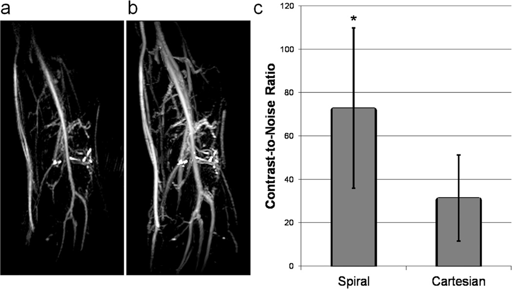
FIG. 6.
Spiral versus Cartesian TSE. a: Spiral TSE; echo spacing = 10 ms. b: Cartesian TSE; echo spacing = 3.8 ms. Confounding venous signal is observed in the Cartesian image and is nearly removed in the spiral image. Additionally, use of the spiral sequence results in better in-plane resolution. c: Quantification of artery/vein CNR based on three regions of interest placed in each of the femoral artery and vein. *P < 0.05.

