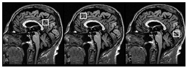
Figure 1. Voxel Locations for Proton MR Spectroscopy.

Three 8 cm3 (2×2×2cm3) voxels are localized on mid-sagittal T1-weighted images. A: Posterior cingulate voxel includes right and left posterior cingulate gyri and inferior precunei. Anterior inferior corner of the voxel is the anterior border of the splenium of the corpus callosum. B: Frontal lobe voxel includes the right and left medial superior frontal gyri. Posterior inferior corner of the voxel is the cingulate sulcus at the level of the anterior margin of the lateral ventricles. C: Occipital lobe voxel includes right and left medial occipital lobes. Anterior superior corner of the voxel is the parieto-occipital sulcus.
