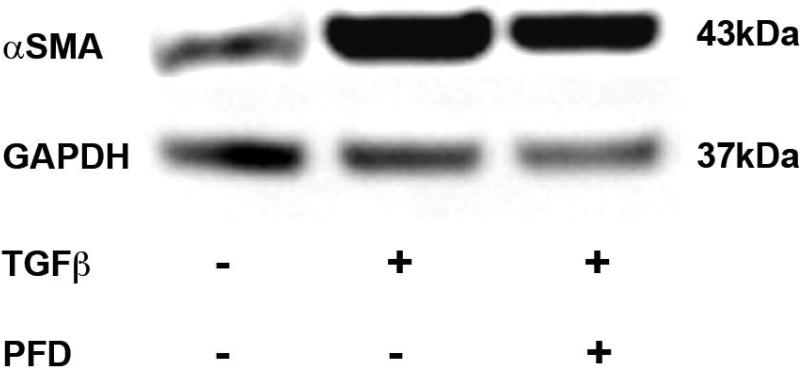
Figure 7.
Immunoblot analysis demonstrating quantitative measurement of αSMA in ECF cultures treated with or without TGFβ1 (1ng/ml) and 200 μg/ml PFD. Equal quantity of protein (30μg) was loaded in each lane. Left lane is untreated control. Cell lysates prepared from TGFβ1-treated cells were loaded in center lane. TGFβ1 + PFD-treated cell lysates were loaded in right lane. GAPDH was used as a housekeeping gene. The 200 μg/ml PFD treatment showed significant decrease in TGFβ1-induced myofibroblast formation. Error bars indicate standard error.
* denotes P < 0.001 (untreated control versus TGFβ1 treatment)
** denotes P < 0.001 (TGFβ1 treatment versus TGFβ1 + PFD treatment).

