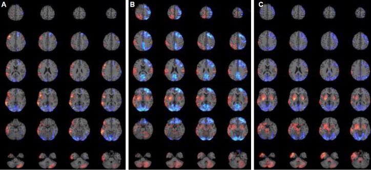
Figure 1.

PET findings of acute phase in three patients with anti-NMDAR encephalitis. Increased metabolism is expressed as red color and decreased metabolism as blue color. (A) Patient 1; increased metabolism in the right frontotemporoparietal regions including the right orbitofrontal cortex, in the right insular cortex, in the bilateral basal ganglia, and in the left cerebellum. Decreased metabolism in the bilateral occipital areas and in the left frontal area. (B) Patient 2; increased metabolism in the right frontotemporoparietal regions including the right orbitofrontal cortex, in the bilateral basal ganglia, in the left parietal cortex, in the bilateral amygdala, in the right insular cortex, in the right thalamus, and in the bilateral cerebellum. Decreased metabolism in the bilateral occipital areas and in the left frontal and parietal areas. (C) Patient 3; increased metabolism in the right frontotemporoparietal regions including the right orbitofrontal cortex, in the bilateral basal ganglia, in the left parietal cortex, in the bilateral amygdala, in the right insular cortex, in the bilateral cerebellum, in the right thalamus, and in the brainstem. Decreased metabolism in the bilateral occipital areas, in the right frontal area, and bilateral parietal areas. PET, positronemission tomography; NMDAR, N-Methyl-D-aspartate receptor.
