
Figure 1.
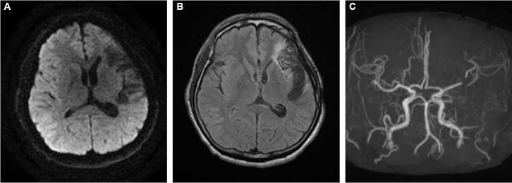
Brain magnetic resonance imaging and angiography at the onset of symptoms. Diffusion-weighted images (A) revealed no acute lesions. Fluid-attenuated inversion recovery images (B) showed old encephalomalacia in left fronto-temporal area and there was a trace of previous clipping of left MCA bifurcation aneurysm on angiography (C). MCA, middle cerebral artery.