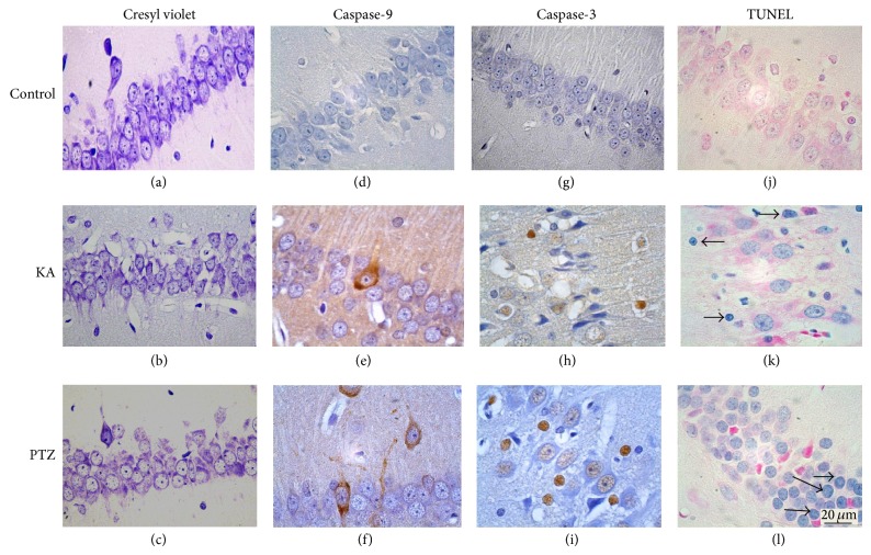
Figure 2.
Representative photomicrographs of hippocampal fields of rats at several times after injection of KA or PTZ. Sections stained with cresyl violet, showing neuronal cells in the hippocampus CA1 field (a, b, and c). Hippocampus showing immunoreactive pyramidal cells to caspase-9 (d, e, and f). Immunoreactive cells to caspase-3. The caspase-3 staining was observed in the cytoplasm and nucleus (g, h, and i). Some pyramidal cells (j and k) and granular cells (l) of dentate gyrus were stained positively for TUNEL (↑).

