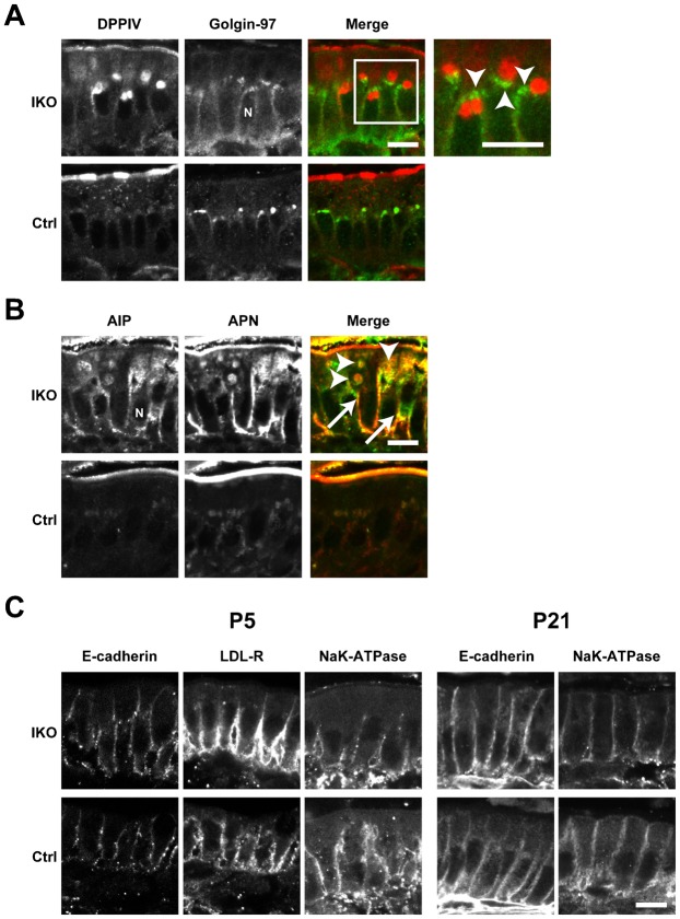
Fig. 3. Localisation of apical and basolateral proteins in the small intestine of Rab11a IKO mice at P21.
(A) DPPIV and the TGN marker Golgin-97 (arrowheads) do not colocalise in intestinal epithelial cells from IKO mice at P21. High-magnification insets (white boxes) are shown to the right of these panels. (B) Localisation of the apical proteins AlP and APN in control (Ctrl) and IKO cells at P21. Merged figures of AlP and APN are shown on the right. Intracellular vacuoles of AlP and APN are indicated by arrowheads. Basolateral localisation is indicated by arrows. (C) Localisation of the basolateral proteins E-cadherin, Na+K+-ATPase, and LDL-R in epithelial cells of the small intestine in Ctrl and IKO mice is not different at P5 and P21. Scale bars: 10 µm. N, nucleus.

