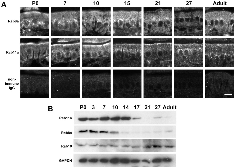
Fig. 7. Localisation of Rab8a and Rab11a and quantification of Rab11a, Rab8a, and Rab10 in the small intestine during postnatal development.
(A) Localisation of Rab8a (top), Rab11a (middle) and non-immune rabbit IgG (bottom), as determined by immunofluorescence, during postnatal (P) days in wild-type epithelial cells of the small intestine. (B) Levels of Rab11a, Rab8a, and Rab10 in the wild-type small intestine during postnatal development. Scale bar: 10 µm.

