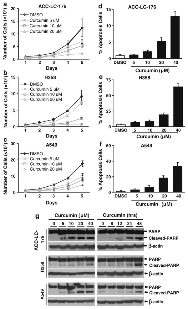
Fig. 1.
Curcumin inhibits cell growth and induces apoptosis of NSCLC cells. NSCLC cell lines ACC-LC-176 (a), H358 (b), and A549 (c) were treated with curcumin with indicated concentrations for 5 days. Cell counting was performed every 24 h using cell counting hemocytometer after treatment (a–c). The percentage of apoptotic cells was analyzed by flow cytometry (d–f) after 24-h treatment. Data were presented as the mean ± SD of triplicate determinations. g NSCLC cells ACC-LC-176, H358, and A549 were treated with curcumin as indicated for 24 h (left panel), or with 10 μM curcumin for different time points (6, 12, 24, 48 h; right panel). Protein lysates from treated cells were used for analyzing the level of PARP and cleaved PARP by western blot analyses

