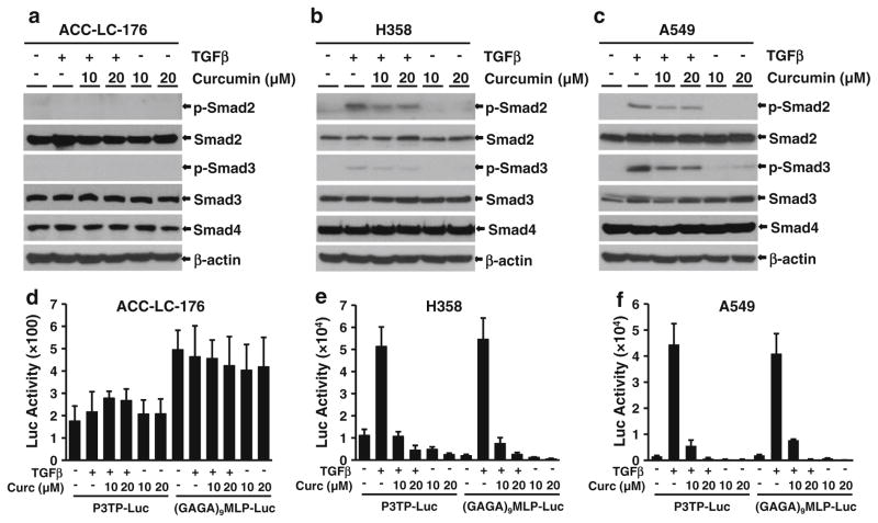
Fig. 6.
Curcumin inhibits TGF-β/Smad signaling in H358 and A549 cells. Western blot analyses were performed using lysates from ACC-LC-176 (a), H358 (b), and A549 (c) cells treated with TGF-β (5 ng/ ml) and 10 or 20 μM curcumin for 22 h. Luciferase reporter assay. ACC-LC-176 (d), H358 (e), and A549 (f) cells were transiently transfected with p3TP-Lux, (GAGA)9 MLP-Luc, and CMV-βgal reporter plasmids, and then pretreated with curcumin of indicated concentrations for 2 h, and 5 ng/ml TGF-β was added thereafter for 22 h. Cell lysates were used to measure luciferase and β-gal activities, and normalized luciferase activities were presented

