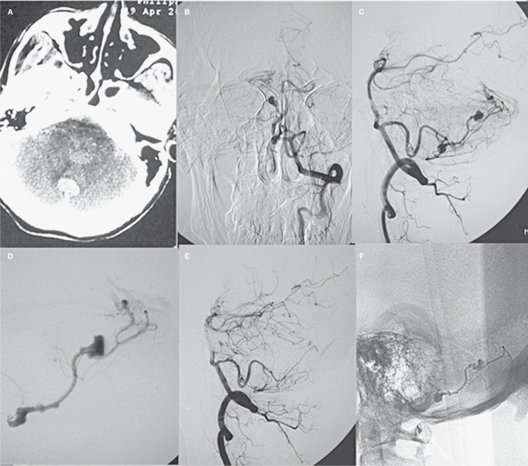
Figure 1.
A middle-aged patient presented with sudden headache. A) CT scan revealed fourth ventricular hemorrhage. B,C) Frontal and lateral view of digital subtraction angiography showed AVM and associated multiple feeding artery aneurysms. D) Super-selective angiography demonstrated associated aneurysms and the AVM. E) Lateral view of the left vertebral artery showed complete obliteration of the AVM and associated aneurysms after embolization. F) Cast of Glubran 2 after the procedure.

