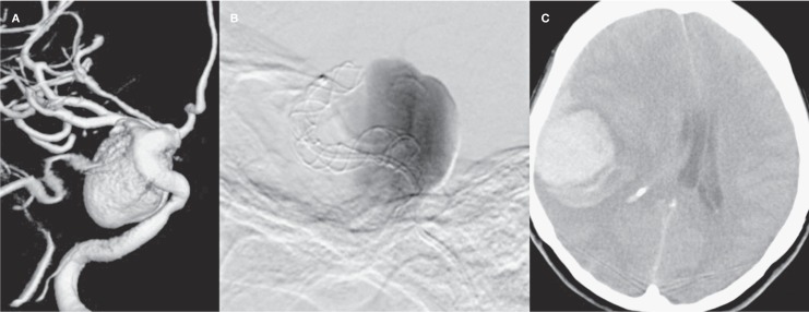
Figure 1.
A) 3D angiography image demonstrating the giant complex aneurysm at right cavernosal segment of ICA of the patient lost due to remote intraparenchymal bleeding within post-procedural 40h. B) DSA image showing wide neck saccular aneurysm with slow flow (Grade 2) during the procedure.C) Native cranial CT axial image demonstrating parenchymal haemorrhage.

