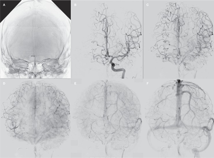
Figure 4.
Test occlusion of the right A1. A) Balloon inflated in the right A1 occluding the origin of the Heubner artery. B-F) Arterial, parenchymal and venous phases of left internal carotid angiogram during test occlusion: abundant leptomeningeal collateral supply over the hemispheres with synchronous venous phase filling.

