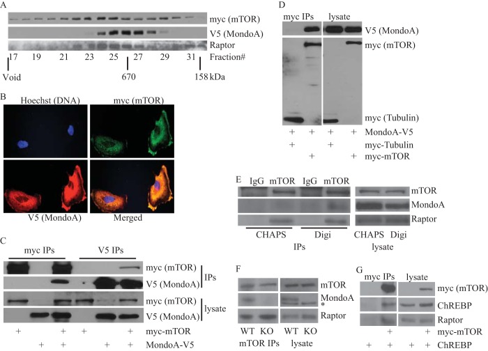
FIG 4.
MondoA and mTOR interact. (A) Lysate from HEK293T cells expressing myc-mTOR and MondoA-V5 was fractionated using a Superose 6 10/300 GL column as described in Materials and Methods. A total of 40 μl from the indicated fractions was subjected to SDS-PAGE, and expression of myc-mTOR, MondoA-V5, and raptor was determined by Western blotting. (B) Representative images showing MondoA-V5 (red) and myc-mTOR (green) subcellular localization in HA1ER cells. (C and D) Levels of indicated proteins in anti-myc or anti-V5 immunoprecipitates and lysate prepared from HEK293T cells transfected with the indicated plasmids were determined by Western blotting. (E) Levels of the indicated proteins in anti-IgG or anti-mTOR immunoprecipitates and lysate prepared from HEK293T cells with the indicated detergents was determined by Western blotting. Digi; digitonin. (F) Levels of the indicated proteins in mTOR immunoprecipitates and lysate prepared from wild-type and MondoA-KO MEF cells were determined by Western blotting (*, nonspecific band). (G) Levels of the indicated proteins in anti-myc immunoprecipitates and lysate prepared from HEK293T cells transfected with indicated plasmids were determined by Western blotting.

