
FIG 5.
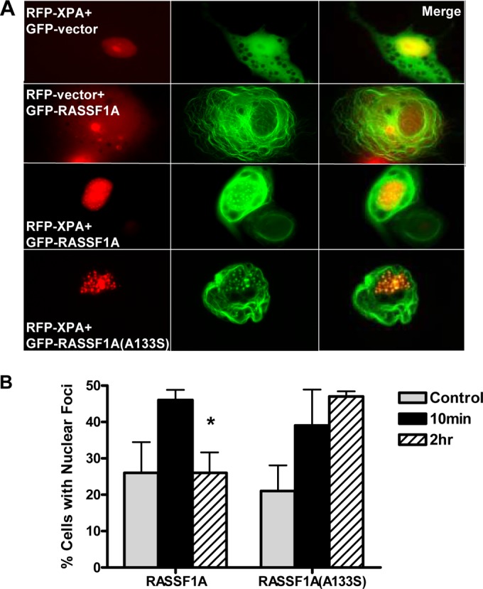
RASSF1A and XPA colocalize in nuclear foci. (A) COS-7 cells were cotransfected with GFP vector or GFP-tagged RASSF1A (GFP-RASSF1A) or the A(133)S SNP variant of RASSF1A and RFP-XPA, and images were captured 24 h later using a fluorescence microscope. Magnification, ×100. (B) The cells were exposed to UV irradiation, and the percentage of cells with XPA and RASSF1A colocalization was quantitated 10 min and 2 h after exposure by randomly selecting 50 cells expressing both GFP-RASSF1A and RFP-tagged XPA (RFP-XPA). Results are expressed as means plus standard deviations (SD) (error bars) from triplicate experiments. The value that is significantly different (P < 0.05) from the value for control cells is indicated by an asterisk.