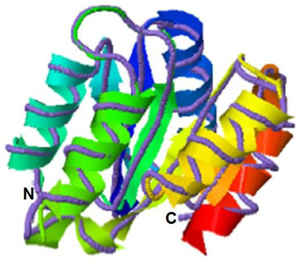
Figure 3.
Model of Cr-CheY structure. The predicted tertiary structure of Cr-CheY (ribbon diagram) shares conserved structural features with the solved structure of a CheY from Helicobacter pylori (purple line, protein database #3H1F). The structural similarities are significant according to I-TASSER, with a TM-score of 0.939 (Zhang, 2008). A TM-score of greater than 0.5 is significant. The N and C-termini are indicated.

