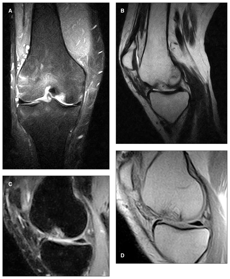
Fig. 3.
Postoperative imaging of MaioRegen implantation. A–B: MRI follow-up at one year. It is possible to note the scaffold in situ and the ongoing differentiation process. C–D: MRI follow-up at two years. The scaffold is still recognizable at subchondral level, but the cartilage layer appears continuous and the subchondral bone shows no signs of edema.

