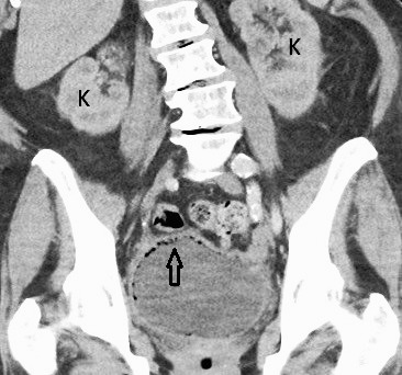
Figure 3.

Contrast-enhanced coronal computed tomography image through the abdomen and pelvis reveals air within the urinary bladder wall (arrow) consistent with emphysematous cystitis. Both kidneys (denoted by K) are visualized with no evidence of perinephric stranding, hydronephrosis, or pyelonephritis.
