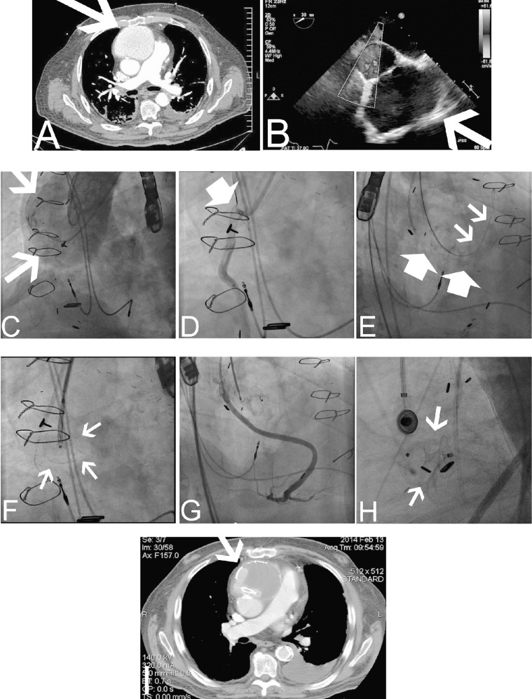
Figure 2.
Endovascular approach to repair an ascending aortic pseudoaneurysm (AAP).
A: Computed tomography angiography (CTA) of the chest exhibits an AAP (arrow) in proximity to the sternum. B: Transesophageal echocardiography (TEE) depicts the shunt between the ascending aorta (AA) and the pseudoaneurysm (arrow). C: Simultaneous contrast injection was performed from a JR4 catheter and pigtail catheter located in the AAP and AA, respectively. Arrows depict the AAP. D: Selective angiography of the venous graft was also performed to assess the distance of the vein graft to the AAP. E: An Amplatz Super Stiff guidewire (small arrows) was advanced via the JR4 catheter into the AAP. A multipurpose catheter was used to engage the nearby vein graft and a 0.014-inch workhorse guidewire was advanced into the graft for protection during the Amplatzer septal occluder device deployment (large arrows). F: The position of the Amplatzer septal occluder delivery system was confirmed in left anterior oblique and right anterior oblique angiographic views and TEE views (arrows). G: Prior to deployment of the device, a vein graft contrast injection was performed to evaluate its patency. H: Final position of the septal occluder device after deployment (arrows). I: Three-month CTA documented stable location of the Amplatzer septal occluder device with evidence of thrombosis in the lumen of the AAP (arrow).

