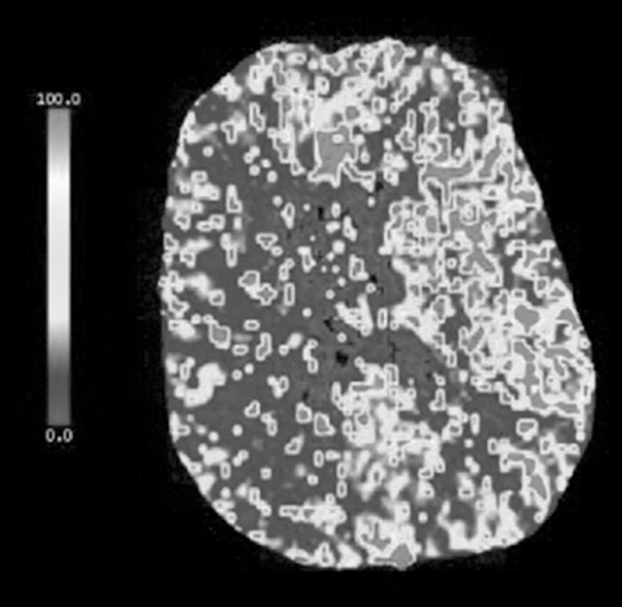
Figure 3.

A computed tomography perfusion scan shows increased mean transit time and decreased blood flow with maintained blood volume involving the majority of the right cerebral hemisphere, suggesting possible ischemic penumbra from slow vascular blood flow. No evidence suggests significant core infarction. There is relative sparing of the right anterior cerebral artery territory likely via collateral flow from the anterior communicating artery.
