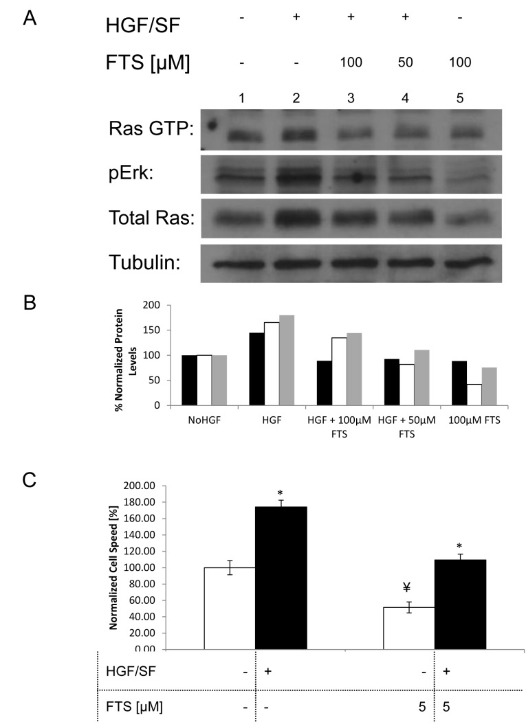
Fig. 1. Effect of Ras inhibition on HGF/SF-induced Ras-ERK signaling and cell motility.
(A), DA3 cells were treated with vehicle (DMSO 0.1% control) or with HGF/SF (80ng/mL), 50 μM FTS+HGF/SF, 100 μM FTS +HGF/SF or 100 μM FTS. After 24h, the cells were lysed and the lysates were subjected to immunoblotting. These results demonstrate that HGF/SF activates Met downstream signaling via the ERK and Ras pathways, while FTS inhibits both basal level signaling and HGF/SF induced signaling via Ras and ERK. The bottom tubulin panel was used as the loading control. (B), Histogram of the protein expression levels, normalized to tubulin. Black bars represents the intensity of Ras GTP in the different samples, white bars represent pERK and gray Bars represent total Ras. (C), a scratch assay was performed by growing DA3 cells to confluency and then scratching the monolayer. Cell media was then exchanged to serum starvation media containing the different treatments. The cells were treated as following: vehicle (DMSO 0.1% control) or with HGF/SF (80ng/mL), 5 μM FTS, 5 μM FTS +HGF/SF. Cell motility rates are compared in the graph that demonstrates that HGF/SF activation of Met increases cell motility (n = 14, P < 0.02 represented by *), while its downstream inhibition by FTS inhibits cell motility (n = 15, P = 0.00025, represented **). Combined treatment with both HGF/SF and FTS leads to reduced cell motility as compared to vehicle treated cells (n = 12, P < 0.02).

