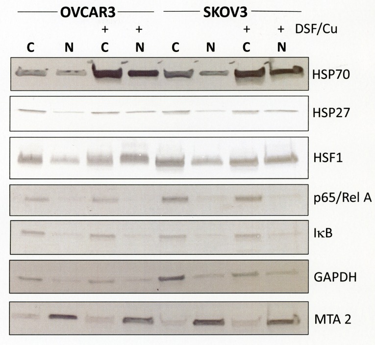
Fig 4. Subcellular distribution of heat shock proteins in response by disulfiram/copper.
OVCAR3 and SKOV3 cells were treated for 8 h with 1 μM disulfiram/copper, separated into a crude cytosolic and nuclear extract as described in the Methods section, and analyzed by Western blot analysis under reducing conditions. GAPDH was used as a cytosolic marker, the transcription factor MTA2 as a nuclear marker.

