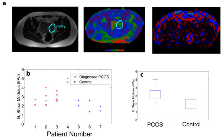
Figure 3.
In vivo magnetic resonance elastography (MRE) studies were performed on seven clinical patients, four of whom had a previous diagnosis of PCOS. (a) T-2 anatomical images were cross-referenced with colorized MR elastograms (0–8 kPa, purple-red respectively) to identify a region of interest (ROI). The ROI was used to measure the shear stiffness, G′ (kPa). (b,c) However, results demonstrate an overall higher stiffness in the ovaries of women diagnosed with PCOS versus age-matched controls. MRE wave images are shown as last color image.

