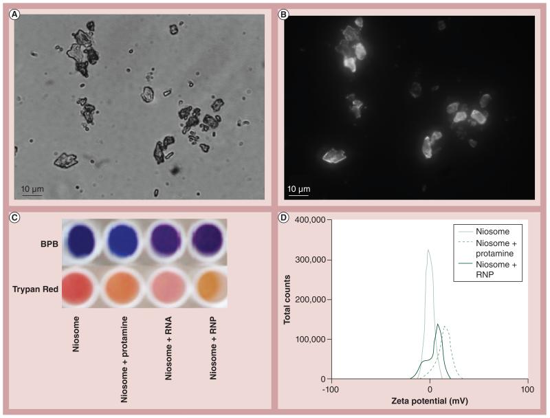
Figure 3. Niosome as a model cell membrane.
Niosomes can be viewed by (A) light microscopy or (B) fluorescence microscopy when BODIPY®–cholesterol was substituted into the membrane. (C) Chromophoric shift of niosome in the presence of protamine, RNA or RNP nanoparticles. (D) Shift in the dynamic light-scattering spectroscopy zeta potential spectrum as a consequence of interaction with protamine or RNP with tripalmitin:cholesterol niosome.
BPB: Bromophenol blue; RNP: RNA:protamine.

