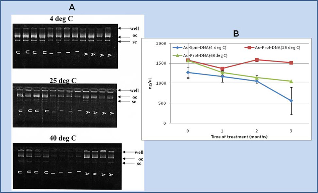
Fig. 3.
Au-spermidine-DNA or Au-protamine-DNA particles, prepared in a 35 mg standard amount [8, 25] were vortex-mixed, sonicated and dried from alcohol, and aliquots of the powders exposed to standard cycles of gamma irradiation (I) and autoclave sterilization (A) as described in the materials and methods; lanes containing untreated DNA are indicated by “U”. The particles were then subjected to exposure at 4 °C, 25 °C, 40 °C, and 60 °C for 2 weeks (panel A) or 1, 2, or 3 months (panel B). Afterwards, the DNA was isolated from 0.5 mg aliquots of the particles with elution buffer and samples were assayed by gel electrophoresis or Hoechst assay as shown above. Supercoiled DNA was completely lost and open circle band staining intensity diminished for Au-spermidine-DNA as shown by gel; however, more DNA was stained by Hoechst assay for Au-protamine-DNA at 25 °C and 60 °C in comparison to Au-spermidine-DNA stored at 4 °C (panel B).

