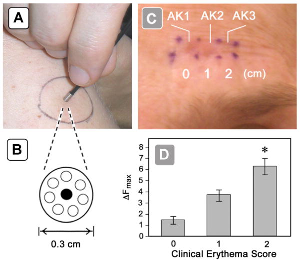
Figure 4.
Clinical use of a non-invasive fluorescence dosimeter for point measurements. (A) Fluorescence probe applied to patient skin during measurement. (B) Close-up of probe tip, viewed end-on. The central optical fiber carries excitation light (405 nm) to the skin, and the peripheral optical fibers carry the fluorescent PpIX emissions back to the detector. (C) Clinical appearance of erythema in 3 actinic keratosis (AK) lesions on a patient’s forehead seen immediately after PDT illumination. Erythema for lesions AK1 and AK2 was graded as a score of 2, while for lesion AK3 the erythema is graded as zero. (D) Post-illumination erythema (x-axis) as a function of the PpIX fluorescence (ΔF) measured prior to treatment (y-axis). (From [49], used with permission.)

