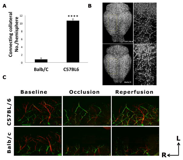
Figure 2. Strain differences in leptomeningeal collateral circulation.
A). The number of connecting collaterals between ACA and MCA is significantly less in the Balb/C compared to C57BL/6 mice (p<0.0001). B). Representative DiI labeling images of Balb/C and C57BL/6 strains at baseline. Magnified views inside of the yellow grids are shown in the right panel. Contrary to the abundant connecting collaterals between MCA and ACA networks seen in the C57BL/6 mice, very few connecting collaterals were found in the Balb/C strain. C). Representative Doppler Optical Coherence Tomography (DOCT) images of MCA and ACA branches at baseline, one hour after MCAO and one hour after reperfusion in C57BL/6 and Balb/C mice. The direction of cerebral blood flow is color-coded, with the blood flowing towards the DOCT probe beam coded as red, and the opposite direction as green. At baseline, the flow direction of MCA and ACA was coded as red and green, respectively. During MCAO, some branches of MCA were filled by ACA retrograde flow via the tortuous anastomoses in the C57BL/6 mouse only, with a reversal of flow direction from red (baseline) to green (occlusion and reperfusion). ACA flow (shown as green) in the Balb/c mice never reached MCA territory during occlusion and reperfusion due to a paucity of the connecting collaterals. The axes indicate the rostral (R) and lateral (L) directions.

