
Figure 3. Cerebral Ischemia induces angiogenesis.
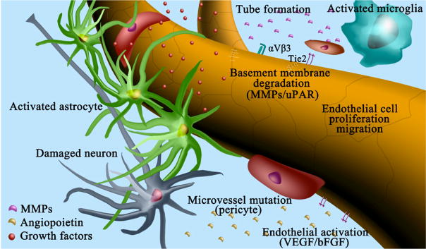
Schematic representation shows the process of angiogenesis in the brain during cerebral ischemia. 1) Damaged neurons, activated endothelial cells and astrocytes release growth factors such as VEGF and bFGF, etc. 2) At the same time, matrix metalloproteinases (MMPs), urokinase-type receptor (uPAR) and inflammatory mediators are highly expressed in the focal environments, which trigger the degradation of basement membrane. 3) Activated endothelial cells proliferate, migrate and sprout into new vessels locally. 4) Activated microglia express many angiogenic and inflammatory factors to promote the formation of new tubes by endothelial cells, endothelial progenitor cells, and mesenchymal stem cells. 5) Upon the activation of Ang/Tie2 signaling, smooth muscle cells and pericytes are attracted to surrounding newly formed microvessels and promote vessel maturation.
 Growth factors,
 MMPs,
MMPs,
 Angiopoietins