Figure 1.
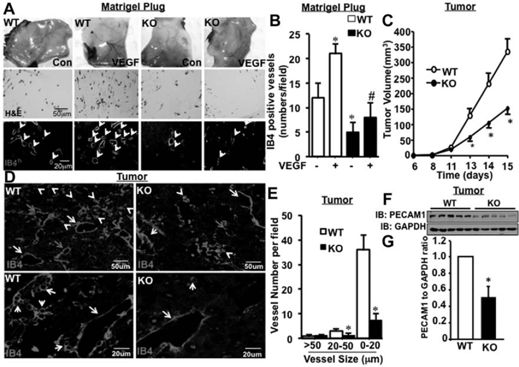
G-protein–coupled receptor-2–interacting protein (GIT1) is required for tumor angiogenesis. A, Matrigel (250 µL) containing vehicle control or vascular endothelial growth factor (VEGF; 50 ng/mL) was injected subcutaneously on the ventral side of the mouse in the groin area. Plugs were isolated 7 days post injection. Cross sections were stained with either hematoxylin and eosin (H&E) or endothelial cell (EC)–specific marker isolectin B4 (IB4) to locate the vessels. B, Vessel density was analyzed by counting the number of IB4-stained vessels per field. *Vs control, #vs control treated with VEGF (n=8; * and #P<0.05). C, Melanoma tumor cells were injected into the thigh muscle of GIT1-wild type (WT; n=5) and GIT1-knockout (KO; n=5) mice. Mean thigh diameters were determined and increased tumor volume was calculated. D and E, Tumors from GIT1-WT and GIT1-KO mice were harvested at day 15 after injection. Tumor tissue sections were stained with IB4 and images were acquired in ×10 and ×40 objective. The number of vessels in 3 different diameter groups (>50 [yellow arrow], >20–≤50 [green arrow], and ≤20 µm [white arrow]) was counted manually using Image Pro Plus software. *Vs WT (n=5; *P<0.05). F and G, Platelet endothelial cell adhesion molecule-1 (PECAM1) expression in tumor samples was detected by Western blot. Quantification of relative expression of PECAM1 normalized to GAPDH. *Vs WT (n=5; *P<0.05).

