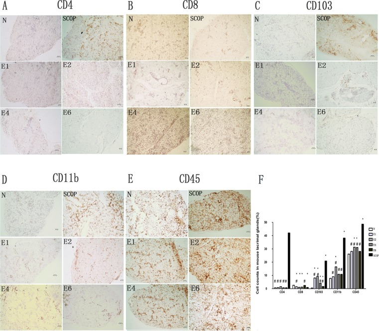
Figure 5. Inflammatory cells infiltration of the Lacrimal Gland.
A-E, immunostained for CD4(A), CD8(B), CD103(C), CD11b(D), CD45(E) in the lacrimal glands sections. F, Cell counts in mouse lacrimal glands stained by immunohistochemistry for CD4, CD8, CD103, CD11b, CD45 sections in the normal group (N), the scopolamine-treated group (SCOP), and after desiccating stress in ICES for 1 week, 2 week, 4 week and 6 week (E1,E2,E4,E6). # P < 0.01 versus the scopolamine-treated group (SCOP), * P<0.05 versus the normal group(N) (Mann-Whitney U test). Original magnification:x20;scale bars = 50 μm. Experiments were repeated three times with two mice per group per experiment.

