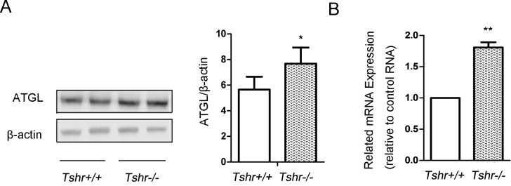
Figure 1. ATGL expression in visceral adipose tissues of Tshr-/- mice increased compared to that of Tshr+/+ mice.

The epididymal adipose tissue was frozen in liquid nitrogen. Protein and mRNA were extracted according to the methods described before. (A) The protein expression levels of ATGL in the white adipose tissues of the Tshr-/- mice and Tshr+/+ mice were detected by Western blotting. The relative ATGL protein levels were quantified by densitometry and normalized with β-actin. (B) The mRNA levels of ATGL in the white adipose tissue of the two types of mice were determined by real-time PCR and normalized with actin. The relative values representing the ATGL mRNA levels in the Tshr-/- mice are reported as fold changes relative to those of the Tshr+/+ mice. The data are from 4 independent experiments and are presented as the mean ± SD. ** p < 0.01 versus Tshr+/+ mice. * p < 0.05 versus Tshr+/+ mice.
