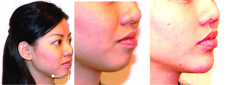
Figure 6.
Mentum augmentation. For augmentation of the mentum, a multi-level approach is generally used. In this case, a cannula was inserted at the pre-jowl sulcus entry point and advanced in both the supraperiosteal layer, as well as the junction between the dermis and subcutis. Augmentation was continued until the patient had a good Steiner Line.39 Left: Before with markings; middle: Before; right: After 1.5mL Radiesse in the mentum.

