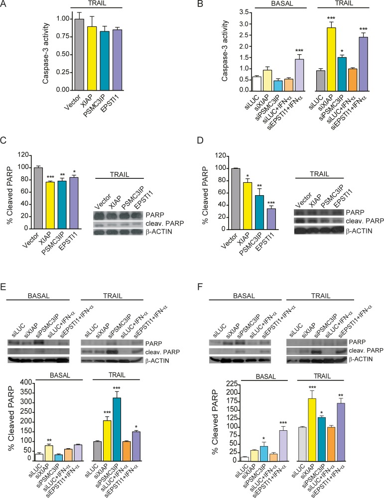
Figure 5. Caspase-3 activity modulation and analysis of cleaved PARP protein levels.
(A) Caspase-3 activity was measured by colorimetric quantification of fluorescent products released from caspase-3 cleaved substrates.in TRAIL-treated MDA-MB-231 cells overexpressing XIAP, PSMC3IP or EPSTI1. None of the overexpressed genes was able to significantly decrease the activity relative to MYC-tag empty transfection vector (Vector) as control. (B) Caspase-3 activity was also measured in MDA-MB-231 cells under basal or TRAIL-treated conditions after gene silencing using specific siRNA targeting XIAP, PSMC3IP or EPSTI1. (C) Immunoblot analysis of cleaved PARP protein levels in gene-overexpressing MDA-MB-231 cells under TRAIL conditions, using MYC-tag transfection vector (Vector) as a negative control, reveals an attenuation of downstream apoptotic cascades. (D) This effect is much more pronounced in TRAIL-treated MCF-7 cells. (E) Analysis of cleaved PARP protein levels after gene silencing in MDA-MB-231 cells. siLUC was used as a negative control. (F) The same analysis in MCF-7 cells shows a more pronounced effect, even under basal conditions. EPSTI1-depleted cells were previously treated with IFN-α at 1000 U/ml for 8h. In apoptosis-induced conditions, MDA-MB-231 and MCF-7 cells were treated with TRAIL for 24h at 80 or 100ng/mL, respectively. XIAP was used as an anti-apoptotic reference in all experiments. Each bar represents the mean ±SD of three experiments performed in duplicate (*P <0.05, **P <0.01, ***P <0.001 vs MYC-tag vector in overexpression assays and vs siLUCIFERASE in silencing).

