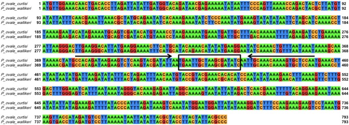
Figure 1. P. ovale reticulocyte binding protein 2 (rbp2) sequence alignment.

The P. ovale curtisi (GU813971) and P. ovale wallikeri (GU813972) rbp2 sequences were aligned using EMBL-EBI Clustal Omega program and visualized in Jalview with the default Jalview nucleotide color scheme (green for adenine, orange for cytosine, red for guanine, and blue for thymine). Primers and probe were designed based on conserved regions between the two P. ovale subspecies. The forward (PoRBP2fwd1) and reverse (PoRBP2rev1) primers are indicated by arrows and the hydrolysis probe (PoRBP2p) binding site (boxed) is located in between the forward and reverse primer.
