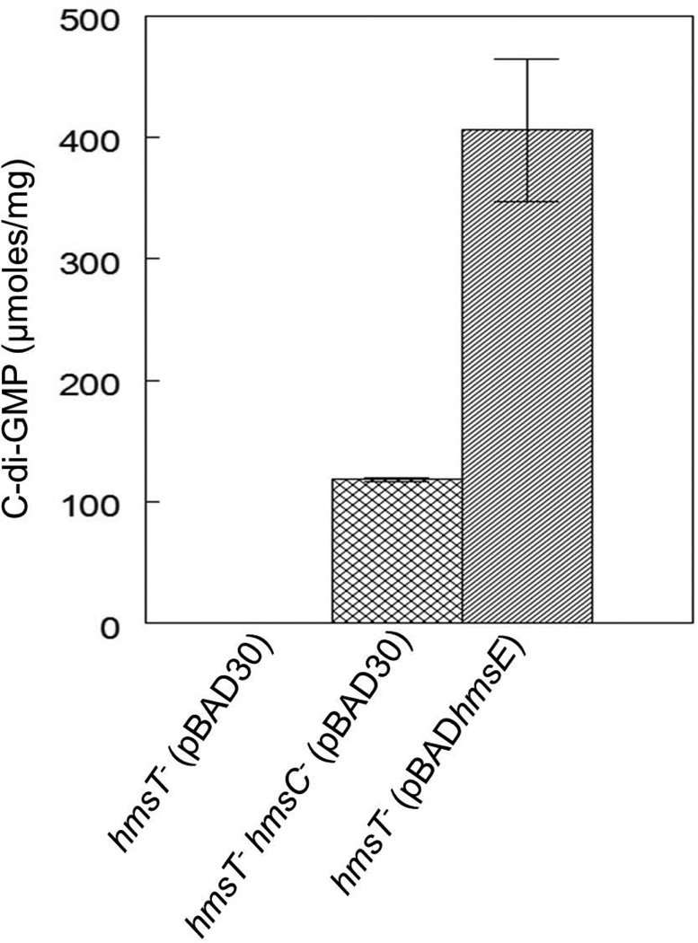
Fig. 3. HmsC and HmsE respectively reduce and increase cellular levels of c-di-GMP through HmsD.
Relative cellular concentrations of c-di-GMP in KIM6-2051+ (hmsT−) and KIM6-2173.3+ (hmsT− hmsC−) carrying either pBAD30 or pBAD-hmsE are shown. Results are from duplicate assays from two independent cultures. Error bars indicate standard deviations. Statistical significance was not calculated because the c-di-GMP concentration in the parent hmsT− strain was below detection limits.

