Figure 2.
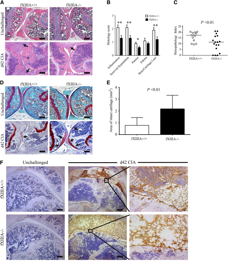
Elimination of fXIIIA results in diminished CIA joint pathology and destruction. (A) Representative hematoxylin and eosin–stained knee joint sections from unchallenged and CIA-challenged fXIIIA+/+ and fXIIIA−/− cohorts. Upon CIA challenge, significant inflammation, synovial hyperplasia (arrows), and erosive pannus (asterisk) are apparent in fXIIIA+/+ mice, whereas knee joints of fXIIIA−/− mice display markedly attenuated pathological features. (B) Semiquantitative microscopic analysis knee joint pathological features from fXIIIA+/+ (n = 13, white bars) and fXIIIA−/− (n = 17, black bars) male mice. Student t test. (C) Scatter plot of composite histopathology index analysis (see Methods) of hematoxylin and eosin–stained knee joint sections. Each symbol represents the composite score for individual mice and bars denote median values for each genotype. (D) Representative safranin-O stained knee joint sections from unchallenged and CIA-challenged fXIIIA+/+ and fXIIIA−/− mice. (E) Quantification of area of intact/preserved articular cartilage per knee based on safranin-O stain. Data are mean ± standard error of the mean with n = 5 mice per genotype and analyzed using Student t test. (F) Representative images of immunohistochemical fibrin(ogen) staining within the knee joints of (left) unchallenged and (middle and right) CIA-challenged mice. Bars represent 200 µm. *P < .05; **P < .01 (B).

