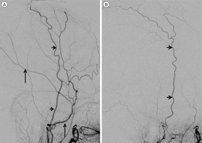
Fig. 2.
Lateral view of the left external carotid artery injection shows the superficial temporal artery (small arrows) with filling of the normal middle meningeal artery (large arrows, A). Lateral view of the right external carotid artery injection shows the superficial temporal artery (small arrows) with no evidence of the middle meningeal artery (B). No tumor blush was seen on either injection.

