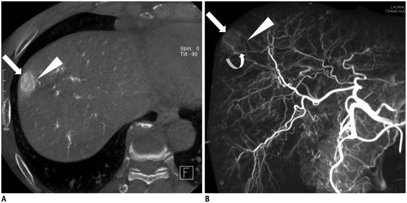
Fig. 1.
60-year-old man with hepatocellular carcinoma.
A. C-arm cone-beam CT shows nodular tumor (arrow) supplied by adjacent subsegmental hepatic artery (arrowhead). B. Maximum intensity projection image of C-arm cone-beam CT obtained at common hepatic artery shows small nodular tumor (arrow) supplied by subsegmental hepatic artery (arrowhead) which was noted on axial image (A). Note another subsegmental hepatic artery (curved arrow) feeding nodular tumor.

