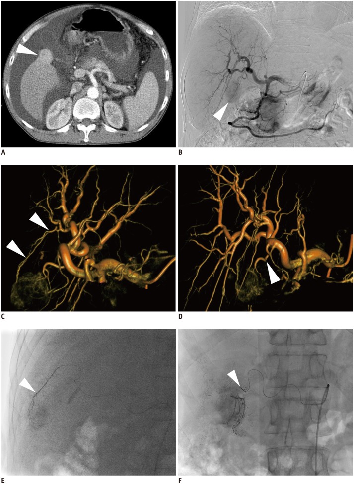
Fig. 4.
47-year-old man with hepatocellular carcinoma and Child-Pugh C class disease.
A. Axial CT scan shows exophytic enhancing nodule (arrowhead) in gallbladder bed. B. Celiac angiography shows tumor staining (arrowhead). C. Volume-rendering image of C-arm cone-beam CT with left anterior oblique projection of 30 degree shows tumor-feeding artery from S5 hepatic artery (arrowheads). D. Volume-rendering image of C-arm cone-beam CT with right anterior oblique projection of 20 degree and cranial oblique projection of 15 degree shows tumor-feeding artery from deep cystic artery (arrowhead). E. Spot image during chemoembolization shows tip (arrowhead) of microcatheter advanced into S5 hepatic artery. F. Spot image during chemoembolization shows tip (arrowhead) of microcatheter advanced into deep cystic artery.

