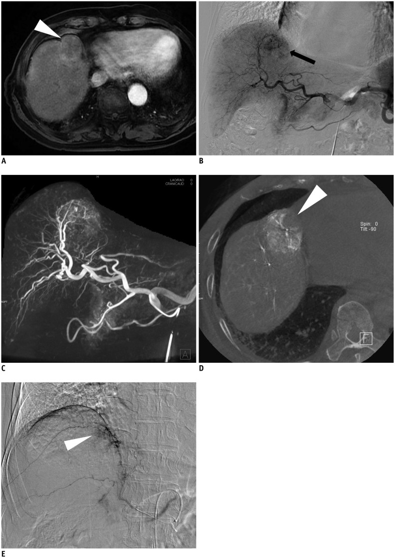
Fig. 5.
78-year-old man with hepatocellular carcinoma.
A. Arterial phase images of gadoxetic acid-enhanced MRI shows exophytic nodule (arrowhead) with faint enhancement. B. Celiac angiography shows hypervascular tumor staining (arrow). C. Maximum-intensity-projection image of C-arm cone-beam CT shows hypervascular tumor staining. D. Axial image of C-arm cone-beam CT shows non-enhancing part (arrowhead) of tumor which suggests presence of extrahepatic collateral artery supplying tumor. E. Angiography of right inferior phrenic artery shows tumor staining (arrowhead).

