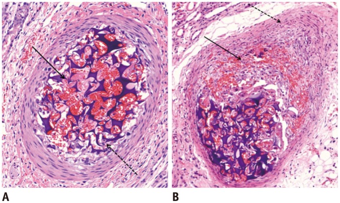
Fig. 1.

Microscopic images of segmental arteries within 1 week after embolization with GSPs.
A. Four days after embolization. There is partially organized thrombus (arrow), and aggregation of macrophages and polymorphonuclear leukocytes between GSPs (dotted arrow). Three layers of vessel wall are preserved. B. One week after embolization. There is focal intimal destruction with transmural inflammation (arrow). Medial and adventitial layers are slightly thickened with proliferation of smooth muscle cells (dotted arrow). Internal elastic lamina is intact (hematoxylin and eosin staining, original magnification × 20). GSPs = gelatin sponge particles
