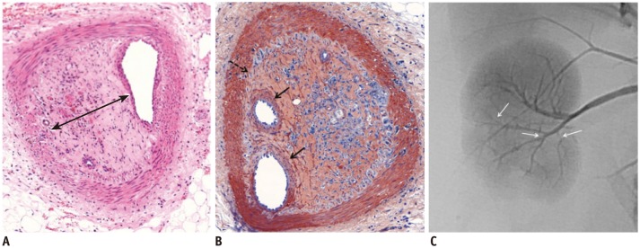
Fig. 3.

Microscopic images of interlobar arteries 3 weeks after embolization with GSPs.
A. Embolized GSPs have completely disappeared, resulting in recanalization. Original lumen is mainly filled with thick organized thrombi (arrow). B. Immunohistochemical staining for smooth muscle actin, showing proliferation of smooth muscle cells in media and thickened intima with migration of smooth muscle cells from media, and interruption of internal elastic lamina (dotted arrow). Migrating smooth muscle cells are mainly distributed around recanalized arterial lumen (arrows). C. Completion angiography, showing diffuse luminal narrowing and multifocal stenosis (arrows) in recanalized arteries of lower pole, compared with non-embolized arteries of upper pole. GSPs = gelatin sponge particles
