
Fig. 4.
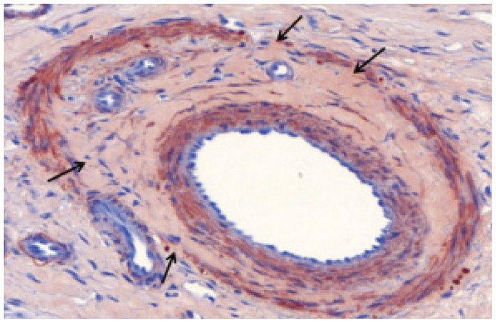
Microscopic image of interlobar artery 4 weeks after embolization with gelatin sponge particles. There are no inflammatory cells. Completely organized thrombus attached to vessel wall, and decreased proliferation of smooth muscle cells in media is observed. Internal elastic lamina has multifocal loss of continuity (arrows) (immunohistochemical staining for smooth muscle actin, original magnification × 20).