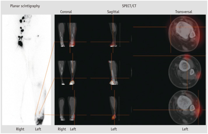
Fig. 3.
65-year-old female patient with clinically swelling of lower left leg suspicious for primary lymphedema. Lymphatic transport disorders (e.g., diffuse distribution of radiopharmaceutical at left lower leg, delayed/missing inguinal lymph nodes of left leg, transport-index 11) were properly detected in planar lymphoscintigraphy (4 hours after injection); due to its three-dimensional imaging options, additionally performed single photon emission computed tomography/CT (SPECT/CT) enables differentiation of anterior versus posterior lymph transport, thus providing accurate anatomic correlation and functional assessment of extent of edema (red colored). Planar lymphoscintigraphy cannot provide these special kinds of morphological information. Physiological lymph transport and distinct visualization of inguinal and iliacal lymph nodes of right leg.

