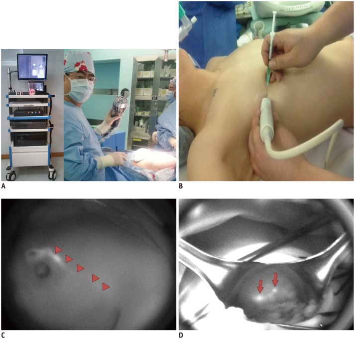
Fig. 2.
Various types of fluorescence imagers can be applied to visualize tissues stained with indocyanine green (ICG) in clinical applications.
A. They range from small, simple, hand-held type to large room-based type. B. ICG solution is injected subcutaneously into periareolar area before operation. C. Lymphatic flows (arrowheads) can be assessed using near infrared fluorescence imager over intact breast skin. D. During operation, intense fluorescence is identified in sentinel lymph node (arrows).

