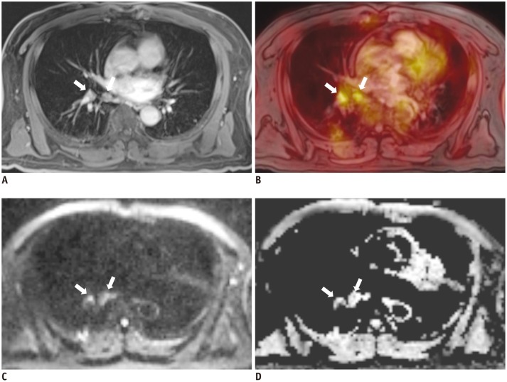
Fig. 5.
66-year-old man with 3.7-cm lung mass (not shown) in right lower lobe.
A. Axial, post-contrast, three-dimensional volumetric interpolated breath-hold examination image showed two small right interlobar lymph nodes (arrows). B. Axial fused FDG-MR/PET image showed increased FDG uptake in interlobar nodes (SUVmax: 4.4 and 3.8). Axial, DWI (b = 400) (C) and ADC map showed diffusion restriction in two lymph nodes (D). Concordant findings of right interlobar lymph nodes on ADC map and on fused MR/PET image increased our confidence in reporting right interlobar lymph-node metastasis, and pathology results confirmed right interlobar lymph-node metastasis. ADC = apparent diffusion coefficient, DWI = diffusion-weighted image, FDG = fluorodeoxyglucose, MR/PET = magnetic resonance imaging/positron emission tomography, SUVmax = maximum standardized uptake value

