Fig. 6.
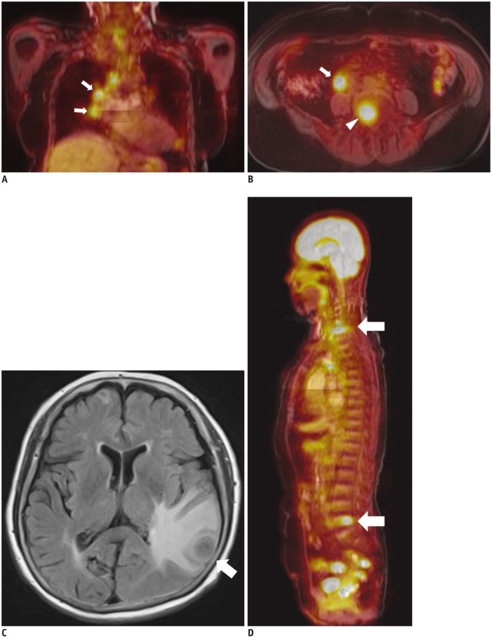
63-year-old male had colon cancer with brain, lung, and mediastinal lymph node (LN) metastasis.
A. Coronal fused FDG-MR/PET image showed mediastinal LNs (arrows) with increased FDG uptake. B. Axial fused FDG-MR/PET image showed metastatic retroperitoneal LN (arrow) and metastatic bone lesion at L5 vertebral body (arrowhead). C. FLAIR image demonstrates brain metastasis in left temporal lobe (arrow). D. Reconstructed sagittal fused FDG-MR/PET image showed bone metastasis in cervical and lumbar spine (arrows). FDG = fluorodeoxyglucose, FLAIR = fluid attenuated inversion recovery, MR/PET = magnetic resonance imaging/positron emission tomography

